Retinal Vascular Occlusion Treatment
Dr. Taylan Öztürk

Retinal Vascular Occlusion Treatment
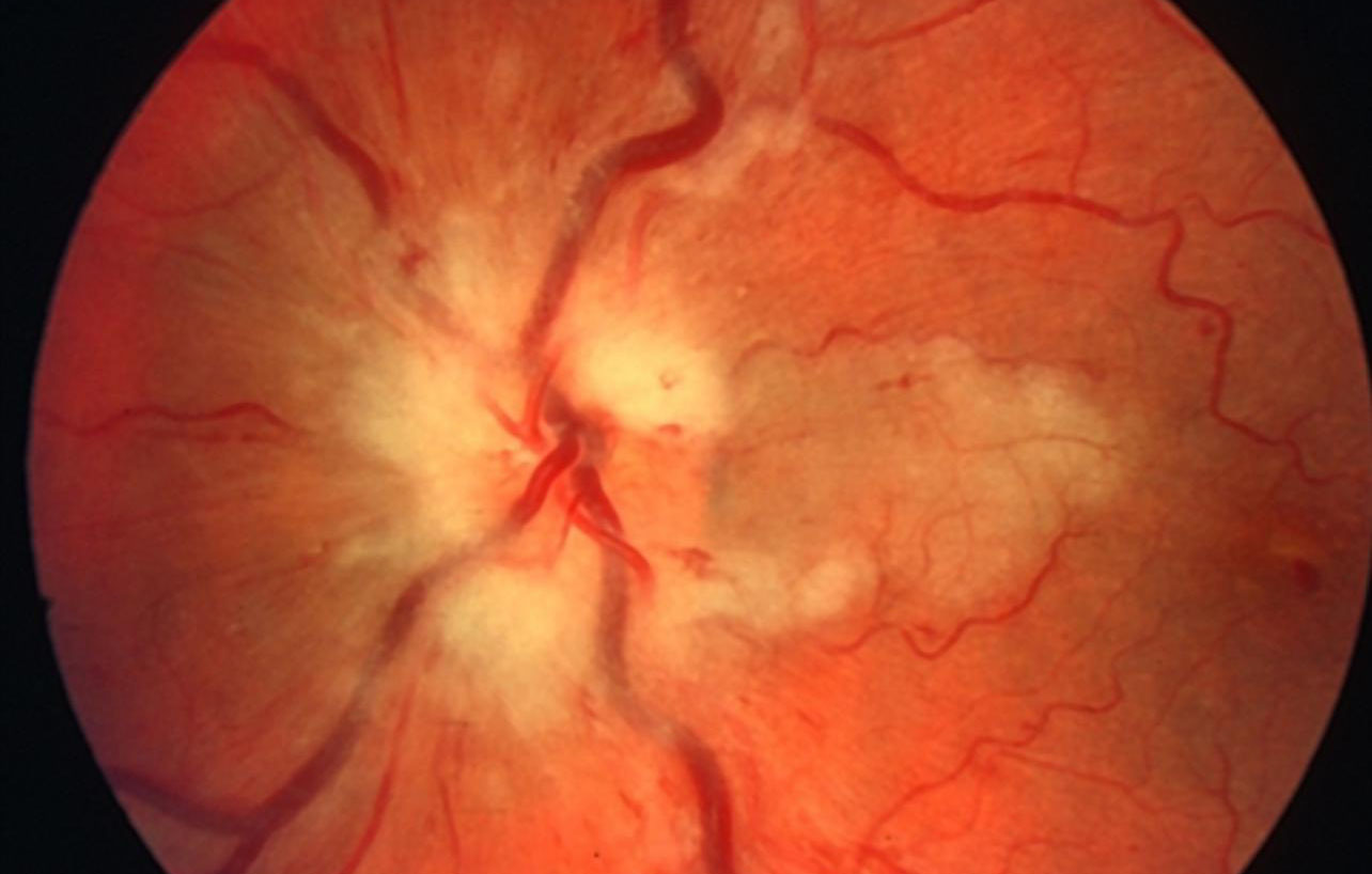
Retinal Vascular Occlusion
Retinal vascular occlusion is a serious eye condition caused by blocked blood vessels in the eye.
Retinal vessels carry blood and oxygen to the retina, which keeps the retina functioning properly. However, if these vessels become blocked, the retina does not receive enough nutrients and oxygen, which can lead to vision loss.
Types of Retinal Vascular Blockages
Central Retinal Vein Occlusion (CRVO): Occurs as a result of blockage of the main vein in the eye and usually causes sudden, painless vision loss.
Branch Retinal Vein Occlusion (BRVO): Occurs when a vein in a part of the retina is blocked and causes blurred vision and visual field damage.
Central Retinal Artery Occlusion (CRAO): Occurs when the main artery in the eye is blocked and can cause sudden vision loss.
Branch Retinal Artery Occlusion (BRAO): Severe visual field loss and loss of vision in a certain area as a result of blockage of the artery supplying a certain part of the retina.


Causes of Retinal Vascular Occlusions
High Blood Pressure
High blood pressure is one of the most common causes of retinal vascular occlusions. High blood pressure can lead to damage to blood vessel walls and blockage of blood vessels.
Diabetes (Diabetes)
Diabetes can disrupt the structure of blood vessels, leading to blockages. Retinal vessels can also be affected, which can lead to vision loss.
High Cholesterol
High cholesterol can cause narrowing and blockage of blood vessels. This is a risk factor for retinal vascular occlusion.
Other Factors
- Smoking: Smoking negatively affects vascular health and increases the risk of blockage.
- Obesity: Excess weight is another factor that impairs vascular health.
- Advancing Age: With age, the elasticity of the vessels decreases and the risk of blockage increases.
Symptoms of Retinal Vascular Occlusions
Sudden Vision Loss
The most common symptom of retinal vascular occlusion is sudden loss of vision. This may vary depending on the severity and location of the blockage.
Loss or blurring of the visual field
Loss or blurring of the visual field is a common symptom, especially in branch retinal vascular occlusions. This occurs when the blockage affects a specific part of the retina.
Pain in the eye
Some patients may experience pain in the eye during or after a retinal vessel occlusion. However, this may not always be the case.

Diagnosis of Retinal Vascular Occlusions
Eye Examination
The diagnosis of retinal vascular blockages is based on a comprehensive eye examination. The ophthalmologist examines the retina with a method called ophthalmoscopy to determine the location and severity of the blockage.
Vision Tests
Vision tests, such as visual field testing and visual acuity testing, are used to assess the impact of the blockage on vision.
Imaging Methods
Fluorescein Angiography: Used to examine blood vessels in the retina.
Optical Coherence Tomography (OCT): Provides detailed images of the retinal layer.
OCTA (Optical Coherence Tomography Angiography): It takes cross-sectional angiographic views of the retinal layers.
Treatment Methods for Retinal Vascular Occlusions
Medication Therapy
In the treatment of retinal vascular occlusions, medications can be used to dissolve blood clots and prevent blockage of blood vessels. Anticoagulants and blood thinners are commonly used medicines for this treatment.
Laser Treatment
Laser treatment is used to destroy abnormal vessels on the retina. This can help prevent further damage to the retina. Laser treatment is usually completed in a short time.
Injection Therapy
Anti-VEGF injections are used to prevent abnormal vessels from forming on the retina and to help existing vessels heal. This treatment can help prevent vision loss.
Intraocular cortisone implant applications aim to rapidly recover the edema in the retina and especially in the yellow spot and improve vision, while providing an effective treatment by reducing intraocular inflammation.
Surgical Intervention
Vitrectomy surgery is performed for intensive intraocular hemorrhages accompanying advanced stage vascular occlusion. While the bleeding intraocular fluid is removed with this surgery, laser treatments and intraocular injections can be performed simultaneously and rapid visual improvement can be achieved in patients.
Retinal Damar Retinal Vascular Occlusion Treatment Process and Price Information
Treatment Process
The treatment of retinal vascular occlusion depends on the severity of the occlusion and the treatment method used:
- Medication: Drug treatment is usually a long-term process and requires regular eye checks.
- Laser Treatment: Laser treatment usually takes 5-20 minutes and does not require hospitalization.
- Injection Treatment: Injections are usually administered once a month and the course of treatment can last for several months.
- Surgical Intervention: Vitrectomy surgery usually takes between 30 minutes and 2 hours and the postoperative recovery period can vary from a few weeks to several months.
Price Information
Prices for the treatment of retinal vascular occlusions vary depending on the treatment method and the patient’s condition.
How Can We Help You with Retinal Vascular Occlusions?
If you are concerned about retinal vascular occlusions or would like more information about treatment options, do not hesitate to contact our team of experts. Schedule an appointment today to protect your eye health and find the most suitable treatment options. Take action now for healthy vision and quality of life!
Protect Your Eye Health! Contact us now to get detailed information about retinal vascular occlusions and get professional support. Take action now for a healthy vision and quality of life!